NT Scan (Nuchal Translucency): Timing, Normal Values and What the Results Mean

The NT scan is done between 11 weeks and 13 weeks 6 days, usually over the abdomen.
For many couples, the NT scan is the first time they see their baby as a real, moving little person. The heartbeat flickers, a tiny hand waves, and for a few minutes everything feels magical. Then the sonologist says something about "the NT measurement" and the mood can shift.
If you've been told your NT is "slightly high" or "borderline," please read this before you start searching. Often, the explanation is simple, and even when it is not, there are clear steps to find out more. This guide explains what the scan is, when it is done, the normal range, what an increased measurement can mean and what comes next. If you want to discuss your own report with a gynaecologist in Noida, details are at the end.
What is the NT scan?
NT stands for nuchal translucency. "Nuchal" means the back of the neck. "Translucency" refers to a small, clear fluid-filled space under the skin at the back of the baby's neck, visible on ultrasound in early pregnancy. All babies have this fluid, and the sonologist measures its thickness.
In babies with some chromosomal conditions (such as Down syndrome), or with certain heart or structural problems, this space tends to be thicker than average. The measurement is therefore a useful marker. But a thicker NT is a sign for further assessment, not a diagnosis.
When is the NT scan done?
The scan is done between 11 weeks and 13 weeks 6 days of pregnancy. At this stage, the baby's crown-rump length (CRL) is between about 45 mm and 84 mm. Outside that window, the fluid space either has not formed fully or has begun to disappear, so the measurement becomes unreliable.
Your doctor will set the date based on your early dating scan, so that you do not miss the window. If you have missed it, there are alternative tests later in pregnancy, such as the quad marker and a detailed anomaly scan.
Who does the NT scan?
This scan needs training and precision. Measurements are taken in millimetres, and a difference of half a millimetre can change the risk estimate. Good practice includes:
- A sonologist trained and certified for NT measurement (for example, with FMF certification, the Fetal Medicine Foundation)
- A correct view: the baby in the mid-sagittal plane (a clean side profile), with the head in a neutral position
- Using the right technique and equipment, with the measurement taken at the widest part of the translucency
Ask whether your sonologist is certified, particularly if your result will guide major decisions.
How is it done?
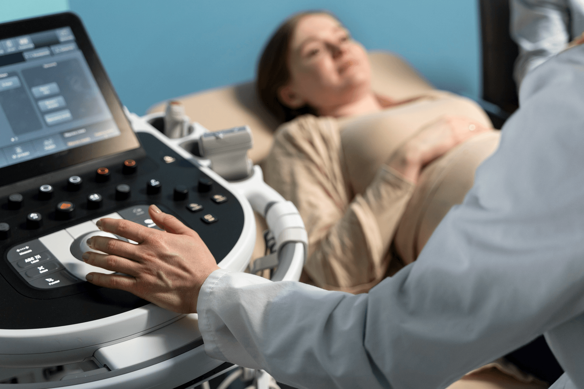
The scan is usually performed over the abdomen with gel and a probe, and takes 15 to 30 minutes. A transvaginal scan may be required if the baby is hard to see. You do not need to fast, and a comfortably full bladder is often requested for a clearer view (follow the instructions of your centre).
The sonologist also checks:
- The baby's heartbeat and number of babies
- Confirming gestational age through CRL
- The baby's overall shape, including head, abdomen and limbs
- The nasal bone, which is present in most healthy babies by this stage
- Blood flow through the heart valve (tricuspid) and the ductus venosus, by Doppler, in expert hands
- The placenta position
- Some centres also look at early structures of the heart and brain
What is a normal NT value?
The thickness is related to the baby's size, so the normal range increases slightly with CRL. As a general rule:
- Less than 3.0 mm (below the 95th centile) is considered normal at most centres. The average NT is around 1.5 to 2 mm.
- 3.0 mm or more is considered increased, and further evaluation is recommended.
- 3.5 mm or more carries a higher risk, and specialist assessment is strongly advised.
In many reports, the NT is stated as a MoM (multiple of the median) for the baby's CRL or is entered into software to calculate risk. A 2.5 mm NT may be normal for a larger CRL and borderline for a smaller one.
An NT measurement is rarely interpreted alone. It is combined with your age, the blood markers from the double marker test (free beta hCG and PAPP-A), and, where available, nasal bone, tricuspid flow and ductus venosus findings to produce a combined risk for trisomy 21 (Down syndrome), trisomy 18 and trisomy 13. See our dedicated article on the double marker test for how to read that report.
How accurate is it?
The combined first trimester screening (NT scan + blood markers + age) detects roughly 85 to 90 percent of Down syndrome pregnancies, with a false-positive rate of about 5 percent. If NT is done alone without the blood tests, the detection rate is lower. It's a screening tool. A "low-risk" result is reassuring but does not exclude all conditions, and a "high-risk" result does not mean the baby is affected.
What can an increased NT mean?
When the NT is increased, there are several possibilities, and most babies with a mildly increased NT turn out well.
- Chromosomal conditions: trisomy 21, 18 and 13, Turner syndrome and others
- Congenital heart defects: an increased NT is associated with heart abnormalities, even when the chromosomes are normal
- Other structural problems: diaphragmatic hernia, abdominal wall defects, skeletal conditions
- Genetic syndromes: a number of rare conditions, such as Noonan syndrome
- Normal babies: many babies with a high NT are healthy at birth. The measurement can be increased temporarily because of delayed lymphatic development
The risk of a problem rises with the thickness of the NT. For example, with an NT of 3 to 3.4 mm, most babies are healthy, but the chance of an issue is higher than average. With an NT above 4 to 5 mm, the chance of a chromosomal or structural issue is considerably higher, though a normal outcome is still possible.
What happens if the NT is increased?
First, don't panic. Understand that you now have more information, and a plan.
- A review of the scan and risk calculation. Your doctor checks the measurement, the CRL, and combines the result with blood markers.
- Counselling. A clear conversation about what the numbers mean, the options and timing.
- Further testing, if appropriate:
- NIPT (non-invasive prenatal testing), a blood test analysing fragments of the baby's DNA in your blood. It is more accurate than the double marker for Down syndrome and carries no risk to the pregnancy. It is a screening test, though, and a positive result should be confirmed.
- Chorionic villus sampling (CVS) at 11 to 14 weeks, or amniocentesis from 15 weeks, give a definitive chromosomal diagnosis. Both carry a small risk of miscarriage. Our article on amniocentesis explains this. For a larger NT, doctors may offer chromosomal microarray in addition to standard testing.
- A detailed anomaly scan at around 14 to 16 weeks by a fetal medicine specialist, and again at 18 to 20 weeks (TIFFA).
- Fetal echocardiography at about 18 to 22 weeks, since heart defects are strongly associated with increased NT, even with normal chromosomes.
- Follow-up for other conditions, such as genetic syndromes, if the NT remains increased or fluid accumulates.
In many cases where chromosome tests are normal and detailed scans (including the heart) are normal, the outlook is good, and the baby's development is usually normal.
What if NT is normal?
A normal NT, along with low-risk blood markers, is reassuring, and the chance of Down syndrome is low. Still, you should have the anomaly scan at 18 to 20 weeks, because the NT scan cannot detect all structural problems and several important conditions develop or become visible later.
Early structural findings in the NT scan
Experienced sonologists increasingly look for major structural problems at the 12-week scan, because some are visible early, for example, absent skull bones (anencephaly), major abdominal wall defects or severe limb problems. This is not a replacement for the 20-week scan, but it can bring important findings forward.
Is the NT scan safe?
Yes. Ultrasound uses sound waves, not radiation, and has been used for decades without evidence of harm to the mother or baby when used appropriately. The scan is non-invasive and painless.
Is it compulsory?
No. Screening is a choice. Some couples decline because the results would not change their decisions. Others want as much information as possible to prepare. You can choose what is right for you, and a good doctor will respect that. What matters is that you understand what the test can and cannot tell you, before deciding.
Legal note on sex determination
In India, it is illegal to disclose the sex of the baby under the PCPNDT Act. Sonologists will not tell you the baby's sex, and prenatal tests are for medical purposes only.
Tips for the day of your scan
- Take your previous reports, including your dating scan and blood test results
- Wear two-piece, comfortable clothing
- Follow your clinic's instructions about bladder filling
- Bring your partner if allowed, as it is a lovely moment to share
- Write down questions in advance
- Keep in mind that the baby's position can affect measurement. If the baby is not lying well, the sonologist may ask you to cough, walk around or return after a short time
Common questions
What if my NT is 2.8 mm? That is below the usual 3.0 mm threshold, but interpretation depends on the CRL. Your doctor will integrate it with other results.
Can NT change in the next scan? The NT disappears naturally after 14 weeks. A later nuchal fold measurement is taken in the second trimester, and a thick fold can also be a soft marker.
Is NT useful in twin pregnancies? Yes, it is measured for each baby, though interpretation is more complex.
Does IVF affect NT results? The blood markers are interpreted differently after IVF. Tell the lab.
If NT is normal, can the baby still have Down syndrome? There's a small chance, which is why screening is not diagnosis.
Is an NT scan the same as a 12-week scan? The 12-week scan is a broader scan. The NT measurement is one part of it.
Plan your antenatal tests with confidence
An NT scan is most useful when it is done at the right time, by a trained sonologist, and explained properly. I look after expectant mothers from Noida, Greater Noida, Ghaziabad and Delhi NCR at Prakash Hospital, Sector 33. To book a consultation, call +91 97023 46853.
Related reading
- Double marker test: normal range and how to read results
- TIFFA anomaly scan at 18 to 20 weeks
- Amniocentesis: when it is advised
- Understanding your ultrasound report
- Pregnancy week by week
This article is for general education. Please discuss your own scan report with your doctor.