Septate Uterus: Miscarriage Risk, Diagnosis and Hysteroscopic Septum Resection
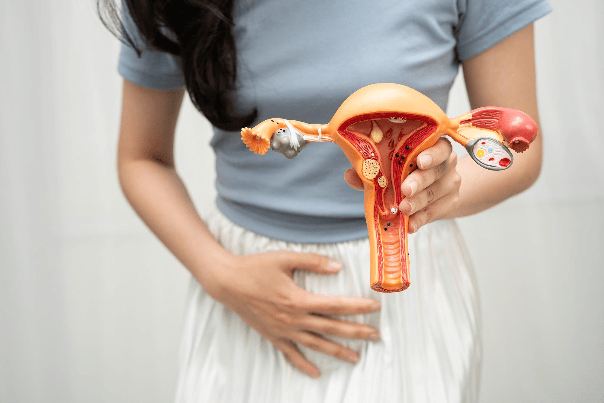
From the outside a septate uterus looks normal; the dividing wall sits inside the cavity.
If you have had a miscarriage, or two, and a scan report now mentions a "septate uterus" or "uterine septum," you probably have two feelings at the same time. One is relief that there may be an explanation. The other is worry about what it means and whether it can be fixed.
A septate uterus is one of the more treatable causes of pregnancy loss, but it is also one of the more frequently misdiagnosed uterine shapes. The decision about treatment depends on getting the diagnosis right and on your own history.
This guide explains what a septate uterus is, how it differs from a bicornuate uterus, how it is diagnosed, how it affects pregnancy, and what the surgery (hysteroscopic septum resection) involves and when it is worth considering. If you are looking for a gynaecologist in Noida with experience in hysteroscopy, details are at the end.
What is a septate uterus?
The uterus forms in a baby girl from two tubes, the Müllerian ducts, which fuse in the middle and then the dividing wall between them dissolves, leaving a single cavity. In a septate uterus, that wall (septum) does not fully dissolve, leaving a band of tissue that partly or completely divides the inside of the uterus into two compartments.
The outside shape of the uterus is normal, with a smooth, convex top. This is the main way to distinguish it from a bicornuate uterus, where the outer contour is indented like a heart.
A septum can be:
- Partial: extends part of the way down the cavity
- Complete: extends all the way to the cervix, sometimes even dividing the cervix or vagina
The septum is made of fibrous or muscular tissue. It has a poor blood supply compared with normal uterine muscle, which is believed to be part of why embryos that implant on it may not grow well.
The septate uterus is the most common uterine anomaly, found in a significant proportion of women with uterine malformations and in a higher share of women with recurrent miscarriage.
Septate versus bicornuate uterus
This distinction is critical, because their treatment and behaviour differ. Our separate guide on bicornuate uterus covers it in depth, but here is the short version.
| Septate uterus | Bicornuate uterus | |
|---|---|---|
| Outside of the top of the uterus | Normal, flat or rounded | Indented, heart-shaped |
| Inside | Wall of tissue dividing the cavity | Two horns of uterine muscle |
| Main pregnancy problem | Early miscarriage | Preterm birth, breech position |
| Treatment | Hysteroscopic resection often considered | Surgery rarely advised |
Misdiagnosing a bicornuate uterus as septate and operating by hysteroscopy would be ineffective and risky. That's why an accurate diagnosis using 3D ultrasound or MRI matters before any surgery.
Does a septate uterus cause symptoms?
Usually not. Periods are typically normal. Many women discover it only after repeated miscarriages, during an infertility work-up, on a routine scan or even during delivery. Some women may notice:
- Recurrent early pregnancy loss
- Second-trimester loss
- Preterm birth
- A baby in breech or transverse position
- Painful periods in some cases
If there is a complete septum with a divided vagina, tampon use may be difficult or intercourse uncomfortable.
How does it affect pregnancy and fertility?
A septate uterus does not stop most women from conceiving, though some studies suggest a modest reduction in fertility. The main effect is on pregnancy outcome.
- Miscarriage. The risk of first-trimester miscarriage is higher than in a normal uterus, and higher than with other uterine anomalies. Some studies report miscarriage in a large fraction of pregnancies, though numbers vary a lot between studies and many women with a septum have successful pregnancies.
- Second-trimester loss and preterm birth. Also more common.
- Malpresentation. Breech or transverse position.
- Fetal growth restriction and placental problems. Slightly increased.
- Caesarean delivery. Higher rates, often due to baby position.
The size and shape of the septum matter. A larger septum, especially one reaching deep into the cavity, tends to cause more trouble. Not everyone with a septum will lose a pregnancy, and not every recurrent miscarriage in a woman with a septum is due to the septum. Other causes, such as chromosomal problems in the embryo, thyroid disease, antiphospholipid syndrome or diabetes, should still be checked.
How is it diagnosed?
2D ultrasound. A normal scan may show a thick lining or a structure in the cavity, and raise suspicion.
3D ultrasound. The most useful and accessible test. It shows the whole uterus from the front, including the outer fundal contour and the shape and length of the septum. With an expert operator, accuracy is high.
Saline infusion sonography (SIS). Fluid is placed in the uterine cavity during ultrasound to outline its shape.
Hysterosalpingography (HSG). X-ray dye test showing the cavity. It displays two cavities but cannot show the outer contour, so it can't separate septate from bicornuate.
MRI. Excellent for the outer contour and the septum structure, particularly in complex cases.
Hysteroscopy. A thin camera passed through the cervix allows direct viewing of the septum. It is both diagnostic and, in the same session, treatment if appropriate.
Laparoscopy. Occasionally combined with hysteroscopy to see the outer surface of the uterus when imaging is unclear.
Because the uterine anomalies often accompany kidney anomalies, a kidney ultrasound is sometimes suggested.
Should a septum be removed?
This is where honest conversation matters, because opinions among experts have changed over time.
Traditionally, many doctors removed the septum in women with recurrent miscarriage, because studies suggested that miscarriage rates dropped after surgery. Those studies were mostly observational, comparing before and after, without a proper control group. More recently, international guidelines have taken a more cautious line. They say that routine removal of a septum in women who have no history of pregnancy loss is not advised, and that the decision should be individualised for women with recurrent pregnancy loss, preterm birth or infertility, after counselling about the uncertain evidence. Trials comparing surgery with waiting are ongoing, and the evidence is still evolving.
In my practice, I consider septum resection when:
- A woman has two or more pregnancy losses (or a late loss or preterm birth) with no other explanation after investigation
- She has unexplained infertility, especially before IVF, and a significant septum
- The septum is large or complete
- She is understanding of the evidence and prefers treatment
I generally do not recommend surgery for a small septum found by chance in a woman who has never had a problem and is yet to try to conceive, without discussion of the pros and cons. There are alternatives, such as watching the pregnancy closely.
Hysteroscopic septum resection (metroplasty)
This is the standard operation. It is performed through the vagina and cervix, with no cuts in the abdomen.
Before surgery. A detailed scan confirms the diagnosis. Surgery is usually timed in the first half of the cycle, when the lining is thin. Some doctors prescribe medicine to thin the lining beforehand.
The procedure. Under anaesthesia (usually short general or spinal), a slim hysteroscope is introduced into the uterus, which is distended with fluid. Using fine scissors, a small electrosurgical instrument or a laser, the surgeon cuts the septum from the bottom upward toward the top, stopping when the septum is gone and the uterine cavity looks symmetrical and the tubal openings are visible on both sides. The surgeon avoids cutting into the thick muscle of the uterine wall. In complicated cases, ultrasound or laparoscopic guidance protects the uterus. The operation usually takes 20 to 40 minutes.
Recovery. It is a day-care procedure for most women. You can go home the same day, with mild cramps and spotting for several days. Many women return to work in a few days.
After surgery. To prevent scar tissue from forming, some surgeons give a course of oestrogen for a few weeks, and occasionally place a balloon or device in the uterus temporarily. A follow-up hysteroscopy or scan after one to two months confirms good healing. Most doctors advise trying to conceive after one or two cycles once healing is confirmed.
Results. Many women report better pregnancy outcomes afterward, though the true size of the benefit is still debated. Reported live birth rates after resection are encouraging in observational studies.
Risks of surgery
Hysteroscopic septum resection is considered safe, but all surgery has risks.
- Uterine perforation. Uncommon, more likely with a thick septum.
- Bleeding
- Infection
- Fluid overload from the distension fluid (a monitored risk)
- Intrauterine adhesions (scarring) that may cause light periods or fertility problems. This is why follow-up matters
- Incomplete resection requiring a second procedure
- Uterine weakness, which might affect delivery planning. A caesarean may be advised if the uterine wall was breached, depending on the findings
- Anaesthesia risks
Your surgeon should go through these before you consent.
Managing pregnancy with a septate uterus
Whether or not surgery is done, once you are pregnant the care should include:
- Early ultrasound to check the pregnancy
- Progesterone support may be considered in selected women with previous losses
- Cervical length checks in mid-pregnancy if there is a history of second-trimester loss or preterm birth
- Growth scans in the third trimester
- Plan for delivery based on the baby's position and other factors. A vaginal delivery is possible if the baby is head-down and other factors are normal
Practical advice for women with recurrent loss
If you have had recurrent miscarriages, a full investigation is wiser than focusing on one cause. It often includes thyroid tests, blood sugar, antiphospholipid antibodies, parental chromosome testing in selected cases, and a uterine assessment. Finding a septum doesn't rule out other causes, and treating only the septum may leave some of the problem unaddressed.
Common questions
Can I get pregnant naturally with a septate uterus? Yes. Most women can conceive. The main concern is carrying the pregnancy.
Is a septum the same as fibroid or polyp? No. A septum is a congenital wall. Fibroids and polyps are growths.
How long after surgery should I wait before trying? Usually one to three months, after healing is confirmed.
Will surgery cure the problem? It improves the shape of the cavity. It doesn't guarantee a successful pregnancy, but it often helps.
Is a caesarean always needed after the surgery? Not always. It depends on the extent of the resection and any complications.
Can a septum be seen on a normal pregnancy scan? Sometimes, but a dedicated 3D scan is more reliable.
Get the right diagnosis first
A septate uterus is a well-recognised and often treatable condition. The most useful step is an accurate diagnosis and a plan built around your history, your age and your goals. I see women from Noida, Greater Noida, Ghaziabad and Delhi NCR at Prakash Hospital, Sector 33. To book a consultation, call +91 97023 46853.
Related reading
- Bicornuate uterus: pregnancy, fertility and delivery
- Recurrent miscarriage: causes and tests
- Hysteroscopy vs laparoscopy
- Asherman's syndrome and uterine adhesions
- Cervical incompetence and cerclage
This article is for general education and is not a substitute for personal medical advice.