Diagnostic Laparoscopy: What It Is, When It's Done, and What It Can Find

Surgeons use slim tools and a camera to perform minimally invasive gynecologic surgery through tiny cuts, working together in a bright, sterile operating room.
There is a specific anxiety that comes with being referred for a diagnostic laparoscopy. You have had symptoms — possibly for months or years — that have not been explained by ultrasound, blood tests, or other investigations. Your doctor has recommended a surgical procedure to look inside and find out what is happening. And now you are left trying to understand what that actually means.
The word "diagnostic" is the important one. A diagnostic laparoscopy is not primarily a treatment — it is an investigation. It uses a tiny camera inserted through a small abdominal incision to directly visualise the pelvic organs in a way that no non-invasive test can match. And in the majority of cases, when a problem is found during the diagnostic procedure, it is treated in the same sitting — so the investigation and the surgery happen together.
Dr. Shachi Singh, consultant gynaecologist and laparoscopic surgeon at Prakash Hospital, Sector 33, Noida, explains what diagnostic laparoscopy involves, who it is recommended for, what it finds, and what happens during and after the procedure.
Why Some Conditions Cannot Be Diagnosed Without Laparoscopy
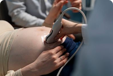
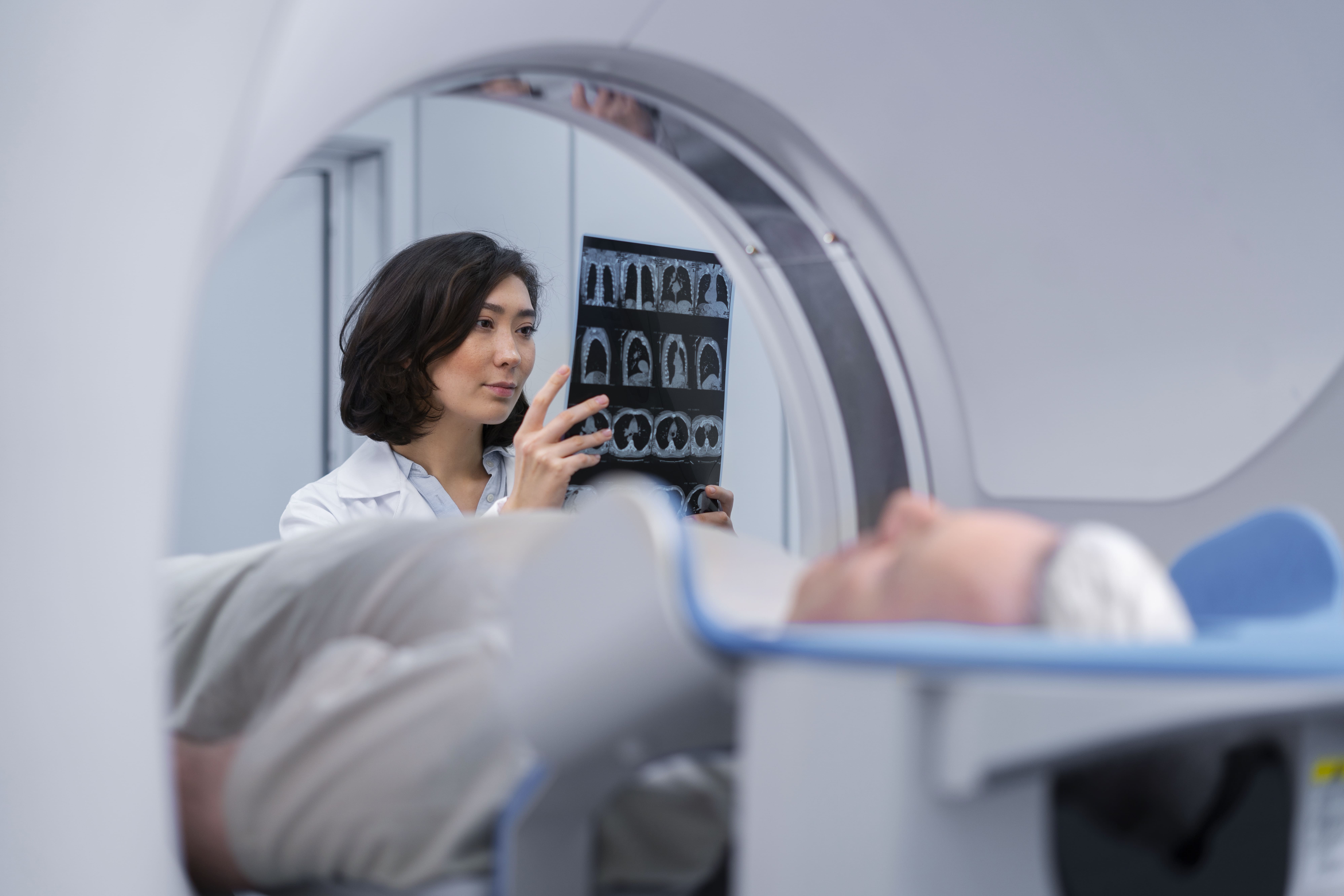
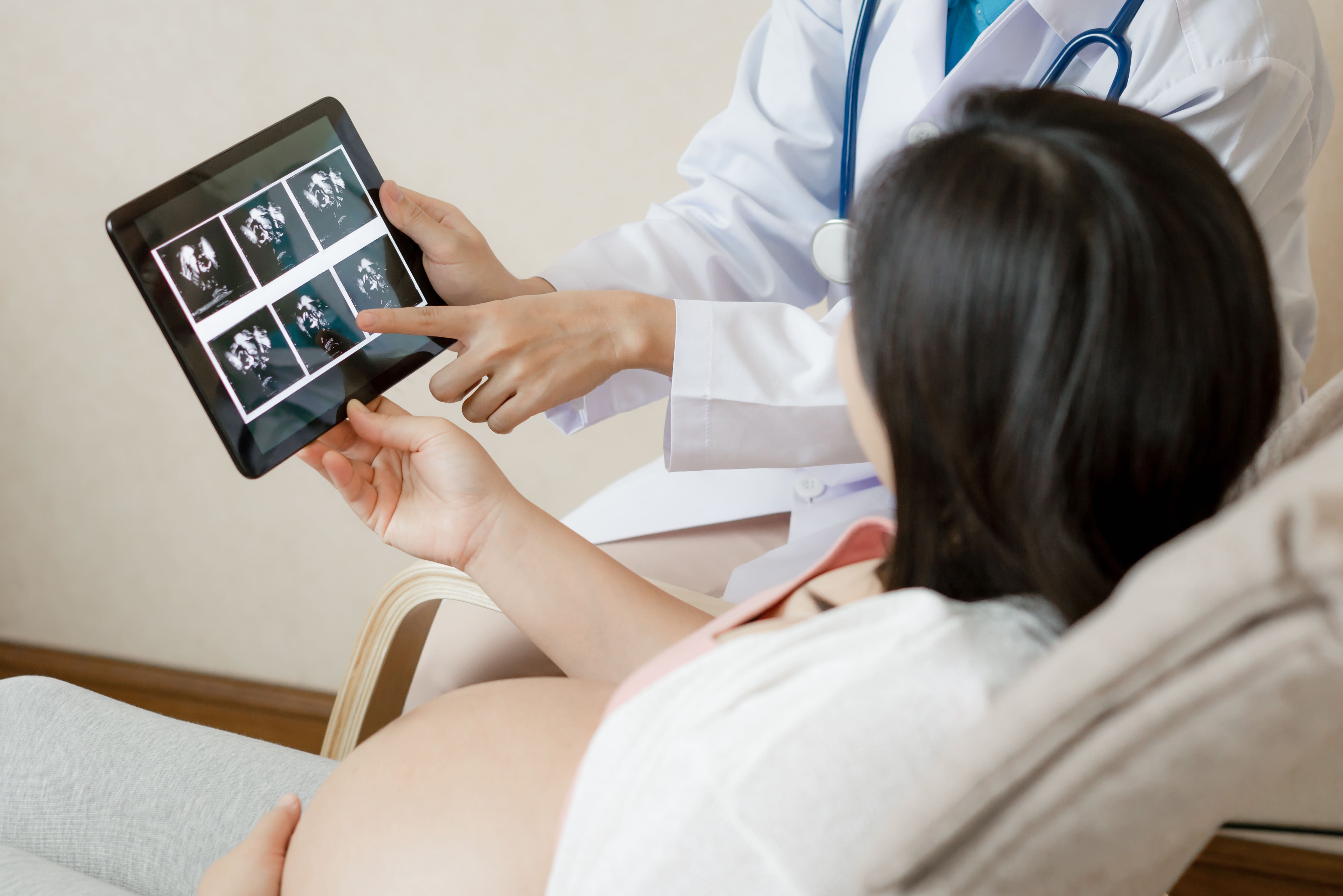
Imaging tests — transvaginal ultrasound, abdominal ultrasound, MRI — are excellent tools but have real limitations. They create pictures of structures. They cannot directly examine the surfaces of the ovaries, the fallopian tubes, the pelvic peritoneum, or the space between organs. They miss fine adhesions between structures. They cannot see small endometriotic deposits on pelvic surfaces. They cannot assess whether a fallopian tube that appears patent on imaging is actually functioning normally.
When these limitations mean a diagnosis cannot be confidently made — or when the diagnosis can be made but treatment requires direct surgical access — diagnostic laparoscopy bridges the gap. It replaces guesswork with a direct, real-time view of the actual anatomy.
When Is Diagnostic Laparoscopy Recommended?
1. Unexplained Pelvic Pain
Chronic pelvic pain that has not been explained by ultrasound and blood tests is one of the most common reasons for diagnostic laparoscopy. The conditions most often found in this setting include endometriosis (which cannot be reliably seen on standard ultrasound), pelvic adhesions from previous infection or surgery, ovarian pathology not fully characterised on imaging, and pelvic inflammatory disease sequelae.
For many women, a diagnostic laparoscopy after years of dismissed or unexplained pelvic pain is the first time a specific cause is identified. The emotional relief of finally having an answer is frequently mentioned by patients as significant — sometimes as important as the treatment itself.
2. Unexplained Infertility
When a woman's standard fertility investigations — hormonal blood tests, ultrasound, HSG for tubal patency — return normal results and yet conception has not occurred, diagnostic laparoscopy is often the next step. It specifically looks for:
- Endometriosis — which affects 25 to 50% of women investigated for infertility and is frequently invisible on standard imaging
- Pelvic adhesions involving the tubes or ovaries
- Tubal blockage or hydrosalpinx not visible or not confirmed on HSG
- Subtle ovarian surface abnormalities
A study from a tertiary care centre in South India found endometriosis in 22.3% of women who underwent laparoscopy for infertility investigation — a significant proportion who would have had no other way to receive that diagnosis.
3. Suspected Endometriosis
Endometriosis affects an estimated 42 million Indian women but remains dramatically underdiagnosed — often by 7 to 14 years from symptom onset. This is because endometriosis cannot be reliably diagnosed by ultrasound or blood tests alone. The definitive diagnosis requires direct visualisation during laparoscopy, with biopsy of suspicious tissue for histological confirmation.
Current international guidelines recommend laparoscopy when endometriosis is clinically suspected and empirical treatment has not provided adequate symptom control.
4. Evaluation of Abnormal HSG Findings
When an HSG (hysterosalpingogram) shows tubal blockage, the finding may represent true obstruction or a false positive from tubal spasm. Laparoscopy with chromopertubation — in which blue dye is injected through the cervix while the surgeon directly observes its passage through the tubes — is the most accurate way to confirm or exclude true tubal blockage.
5. Following Failed Fertility Treatments
When IUI or other fertility treatments have failed without a clear explanation, diagnostic laparoscopy may be recommended to exclude structural pelvic pathology before proceeding to IVF. In some cases, IVF success is significantly improved by first treating conditions found during laparoscopy — particularly endometriosis and adhesions.
6. Other Indications
- Evaluation of suspected pelvic inflammatory disease not responding to antibiotics
- Assessment of pelvic masses not fully characterised on imaging
- Second-look procedure after previous surgery to assess for adhesion reformation
- Evaluation of uterine anomalies (in combination with hysteroscopy)
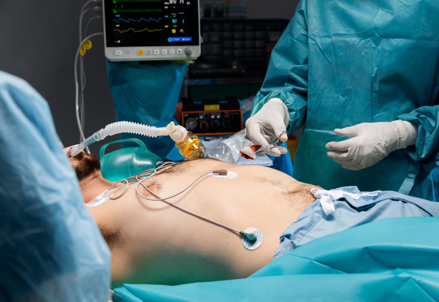
What Happens During Diagnostic Laparoscopy
Anaesthesia: General anaesthesia. You are completely asleep for the duration.
Setting up: Three small incisions are made — typically one at the navel (for the laparoscope/camera) and one or two additional incisions in the lower abdomen for grasping instruments.
Insufflation: The abdomen is gently inflated with carbon dioxide gas, lifting the abdominal wall away from the organs and creating a working space with a clear view.
Systematic inspection: The surgeon examines the pelvic organs methodically. The uterus — its surface, shape, and mobility. Both ovaries — their size, surface, presence of cysts or deposits. Both fallopian tubes — their course, fimbriae, and mobility. The pelvic peritoneum (the lining of the pelvic cavity) — looking for endometriotic deposits, adhesions, or inflammatory changes. The Pouch of Douglas (the space between the uterus and rectum) — a common site for endometriosis.
Chromopertubation (tubal dye test): If tubal patency assessment is part of the procedure, methylene blue dye is injected through the cervix while the surgeon watches for it to spill from the fimbriated ends of the tubes. This directly confirms whether each tube is open.
Documentation: Everything found is documented — often with intraoperative photographs or video. This provides a permanent record and informs post-operative management planning.
Diagnostic and Operative — Usually in the Same Procedure
This is the aspect of diagnostic laparoscopy that many patients do not know before their surgery: when a treatable condition is found, treatment typically happens in the same procedure.
If endometriosis deposits are found, they can be excised or ablated. If adhesions are identified, they can be cut and released (adhesiolysis). If an ovarian cyst is seen, it can be removed. If the dye test identifies a proximal tubal obstruction amenable to cannulation, this can be attempted. If pelvic inflammatory disease sequelae are found, washout and assessment can be performed.
The ability to move seamlessly from diagnosis to treatment — without requiring a second anaesthetic and a second surgical procedure — is one of the most practical advantages of laparoscopy over any non-invasive diagnostic approach.
When a woman agrees to diagnostic laparoscopy, she should understand and consent to the possibility that operative intervention will happen in the same sitting. This should be discussed and documented in the pre-operative consent process.
What Diagnostic Laparoscopy Finds — and How Often
In studies of women undergoing diagnostic laparoscopy for infertility or pelvic pain, the most common findings are:
- Endometriosis: Found in 22 to 50% of women investigated for infertility; up to 70% of women with chronic pelvic pain
- Pelvic adhesions: Particularly in women with a history of pelvic infection, appendicitis, or previous surgery
- Ovarian cysts or endometriomas: Particularly in women with PCOS or endometriosis
- Tubal pathology: Hydrosalpinx, distal tubal occlusion, peritubal adhesions
- Normal pelvis: In approximately 15 to 25% of cases, no specific pathology is found — which, while not providing a treatment target, confirms that significant structural pelvic disease is not present and informs the next steps in management
Recovery After Diagnostic Laparoscopy
Pure diagnostic laparoscopy — where only inspection was performed without significant treatment — has the shortest recovery of any laparoscopic procedure. Most women are discharged the same day.
Day of surgery: Grogginess from anaesthesia, mild incision site soreness, the characteristic CO2 shoulder discomfort (resolves in 24 to 48 hours). Light diet and rest.
Days 1 to 3: Light activity at home. Mild soreness improving daily. Most women return to light desk work by day 2 to 4.
If treatment was performed in the same procedure: Recovery depends on what was done — adhesiolysis or minor endometriosis excision adds minimal recovery time. Larger endometrioma removal or extensive adhesiolysis will lengthen recovery similarly to those specific procedures.
Return to normal: Typically 5 to 7 days after a purely diagnostic procedure. Longer if operative intervention was performed.
Laparoscopic Gynaecological Care in Noida and Greater Noida
Dr. Shachi Singh at Prakash Hospital, Sector 33, Noida, performs diagnostic and operative laparoscopy for women across Noida and Greater Noida — for unexplained pelvic pain, infertility investigation, suspected endometriosis, and tubal assessment.
If you have been told you may need a diagnostic laparoscopy, or if you have had symptoms that have not yet been explained, a proper consultation is the right starting point.
To book a consultation with Dr. Shachi Singh, call: +91 97023 46853
Clinic Hours: Monday to Saturday, 9 AM – 6 PM | Sunday, 10 AM – 2 PM
Clinic Address: D-12A, 12B, Sector-33, G.B. Nagar, Noida, Uttar Pradesh 201301
Frequently Asked Questions
1. Will I be treated during the diagnostic laparoscopy or only diagnosed?
In most cases, if a treatable condition is found during the diagnostic procedure, it will be treated in the same sitting — without requiring a second surgery. This is one of the key advantages of laparoscopy. You should be informed and consent to this possibility before your procedure.
2. Is diagnostic laparoscopy painful?
The procedure is done under general anaesthesia — you feel nothing during it. Afterwards, most women experience mild incision site soreness and temporary shoulder discomfort from residual CO2 gas. This is significantly milder than most people anticipate.
3. How long does a diagnostic laparoscopy take?
A purely diagnostic procedure typically takes 20 to 45 minutes. If treatment is performed simultaneously — endometriosis excision, adhesiolysis, cyst removal — operating time increases depending on what is found and what is done.
4. What if nothing is found during the laparoscopy?
A normal laparoscopy is a meaningful result — it confirms the absence of significant pelvic structural pathology. The management plan then shifts: if the indication was infertility, IVF may be the appropriate next step. If the indication was pelvic pain, other causes are investigated.
5. Can diagnostic laparoscopy be combined with hysteroscopy?
Yes — it is common to perform diagnostic laparoscopy and hysteroscopy together when both the external pelvic anatomy and the internal uterine cavity need to be assessed. Hysteroscopy uses a camera through the cervix to view the uterine cavity without any abdominal incisions.
This blog is written for educational and informational purposes only. Please consult Dr. Shachi Singh or a qualified gynaecological surgeon for an assessment specific to your symptoms and clinical situation.