TIFFA Scan (Anomaly Scan) in Pregnancy: What It Checks at 18 to 20 Weeks

The TIFFA scan is best done at 18 to 20 weeks and usually takes 30 to 45 minutes.
Somewhere around the fifth month, your doctor says, "Time for the TIFFA scan." Most women nod, book the appointment and wonder privately what exactly they are signing up for. Is it just another ultrasound? Is it the one where something might be found?
It is the most detailed scan of your pregnancy, and for many parents it is also the most emotional. In this guide I will explain what the scan is, what the sonologist looks at, what it can and cannot detect, how to prepare and what happens if the result is not perfectly clear. If you want to discuss your own reports with a gynaecologist in Noida, you will find details at the end.
What does TIFFA stand for?
TIFFA stands for Targeted Imaging for Fetal Anomalies. It is also called the anomaly scan, level 2 ultrasound, detailed fetal scan or 20-week scan. In plain terms, it is a very careful, organ-by-organ ultrasound check of your baby, your placenta and your amniotic fluid.
It is not a routine growth scan. Time is taken to look at the structure of each organ system, and the sonologist usually has special training and high-resolution equipment.
When is it done?
The best window is 18 to 20 weeks, and it can be performed anywhere from 18 to 22 weeks. Earlier than 18 weeks, the organs are too small to examine fully. Much later, the baby may be positioned so that measurements are harder, and any decision-making is more limited.
In India, the TIFFA scan is typically done between 18 and 20 weeks 6 days. Your doctor will tell you the exact date based on your dating scan.
What does the scan check?
A good TIFFA report covers a long list. The sonologist works through it one system at a time.
Head and brain. The shape of the skull, the brain's fluid spaces (ventricles), the cerebellum and the midline structures. This helps detect problems such as hydrocephalus or absent brain structures.
Face. The upper lip and palate, the nose, eyes and profile. A cleft lip can often be seen. A cleft palate is harder to detect.
Spine. Each vertebra, plus the skin over the back. This looks for neural tube defects such as spina bifida.
Neck and chest. The shape of the chest, the lungs and the diaphragm. The scan can pick up a diaphragmatic hernia or masses in the lungs.
Heart. The four chambers, the connections to the main vessels (the outflow tracts), the heart rate and rhythm. Heart defects are among the commonest birth defects, and some cannot be seen at this stage, which is why a dedicated fetal echocardiography is advised in higher-risk pregnancies.
Abdomen. The stomach bubble, the bowel, the liver, the kidneys and the bladder. The wall of the abdomen is checked for openings.
Limbs. Arms, legs, hands and feet, including the number of bones and the position of the joints.
Umbilical cord. The number of vessels (normally three: two arteries and one vein) and where the cord attaches to the placenta.
Placenta. Its position (to rule out placenta previa), its thickness and its texture.
Amniotic fluid. Too much (polyhydramnios) or too little (oligohydramnios) can point to problems.
Fetal growth. Head circumference, abdominal circumference and femur length are measured to estimate weight and check growth against your gestational age.
Cervical length. Many centres measure it with a transvaginal scan if there is a history of preterm birth or a short cervix suspicion. A short cervix can be treated.
Uterine artery Doppler. Sometimes used to estimate the risk of preeclampsia and growth restriction.
What can the scan detect, and what can't it?
A properly performed anomaly scan picks up a large share of major structural abnormalities. Detection rates vary widely depending on the type of defect:
- Neural tube defects and abdominal wall defects: high detection rates
- Major brain, kidney and limb abnormalities: good detection rates
- Heart defects: moderate to good, depending on the defect and the skill of the operator
- Subtle problems, like small holes in the heart (VSD), minor kidney issues or some cleft palates: often missed
It is important to be honest about the limits. No scan finds everything. Some conditions, such as many genetic syndromes, deafness, autism, mild heart rhythm problems, some metabolic diseases and cerebral palsy, cannot be seen on ultrasound. Some structural problems develop later in pregnancy and were simply not present at 20 weeks.
The quality of a scan depends on:
- The skill of the sonologist. This matters more than the machine.
- The baby's position. If the baby is facing the wrong way, some structures cannot be viewed.
- Maternal body mass index and abdominal scarring. Extra tissue reduces image clarity.
- The amount of amniotic fluid.
- Twin or multiple pregnancy, which takes longer and is harder.
If the baby is lying in an awkward position, the sonologist may ask you to walk around, empty your bladder or return in a week to complete the picture. A repeat scan does not mean something is wrong. It usually means a few images were unclear.
How is the scan done?
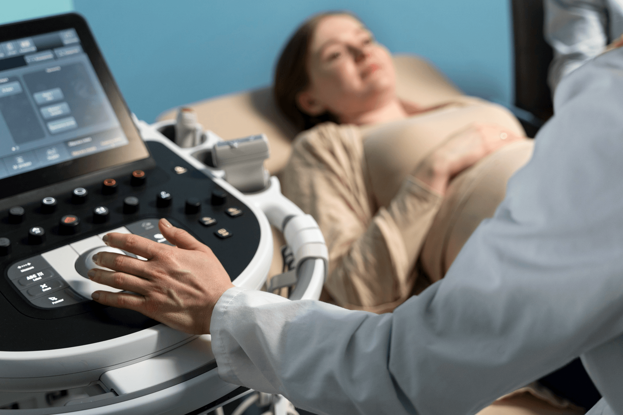
You lie down on a couch and gel is applied to your abdomen. The sonologist moves a probe slowly across your belly. The room is usually dimmed. The scan takes 30 to 45 minutes, sometimes longer if the baby is active or in a difficult position. Occasionally a transvaginal scan is used as well.
You can usually bring your partner. Some centres allow other family members, though policies differ.
How to prepare
- Wear comfortable, two-piece clothing so the abdomen is easy to expose.
- A full bladder is not usually needed at 18 to 20 weeks, though some centres ask for a comfortably full bladder for better images. Follow the instructions you are given.
- Bring your previous reports, including your dating scan, NT scan and double marker results.
- Eat normally. A snack beforehand sometimes makes the baby more active.
- Keep your questions ready. It is a good time to ask about placenta position, fluid and growth.
A word on the baby's sex
Many parents hope to learn the sex of the baby at this scan. In India, disclosing the sex of the fetus is prohibited under the PCPNDT Act. Sonologists are not allowed to tell you, and no ethical clinic will. The scan is for your baby's health, and that is the focus.
Understanding your report
A normal report says there are "no obvious structural abnormalities detected." That is reassuring. Note the wording: "no obvious" and "detected." It means the scan did not see anything wrong, not that guarantees were given.
Reports commonly list some soft markers and minor findings. These are small variations that are usually harmless but are looked at in context.
- Choroid plexus cyst: a tiny fluid pocket in the brain, usually disappears by itself
- Echogenic intracardiac focus (EIF): a bright spot in the heart, usually of no consequence on its own
- Mild pyelectasis: slight widening of the kidney drainage space, rechecked later
- Single umbilical artery: a cord with two vessels instead of three, which needs growth monitoring
- Low-lying placenta: often moves upward as the uterus grows, and is rechecked at 32 weeks
Your doctor will explain what each one means in your case, together with your double marker risk and other results.
What if something looks unusual?
Hearing that the sonologist saw "something" is frightening. Here is the usual sequence.
- Don't panic and don't search at midnight. Many suspicious findings turn out to be nothing, or mild and treatable.
- A repeat scan or review by a fetal medicine specialist, often within one to two weeks, provides a second opinion with more detailed equipment.
- Additional tests: fetal echocardiography for heart concerns, a fetal MRI for brain or chest concerns, and blood tests for infection or genetic conditions.
- Diagnostic testing like amniocentesis may be offered if there's a suspicion of a chromosomal or genetic cause.
- Counselling and planning. Some conditions are treated before or soon after birth, some need delivery in a hospital with a neonatal intensive care unit, and some require sensitive discussions about the outcome. Whatever the finding, you are entitled to full information and unhurried support. In India, the law (the MTP Act) allows termination beyond 20 weeks for substantial fetal abnormalities after assessment by a medical board, and your doctor can explain how this works.
Many babies with a diagnosed problem go on to do very well, particularly where birth is planned in the right centre with the right team.
Is the scan safe?
Yes. Ultrasound uses sound waves, not radiation. Decades of use have found no harm to mothers or babies at diagnostic levels. Scans should be done for medical reasons by trained professionals and not repeated for entertainment, since the machine settings can raise tissue temperature slightly with long exposure.
Questions I get asked often
Do I need the TIFFA scan if my earlier scans were normal? Yes. The NT scan at 12 weeks checks chromosomal risk markers, while the anomaly scan looks at the structure of the organs, which are better developed by 20 weeks.
Can the scan diagnose Down syndrome? Not definitively. It can show soft markers or structural features linked to chromosomal conditions, but a diagnosis requires a test like amniocentesis.
What if I missed the 18 to 20 week window? Go as soon as possible. A scan at 22 to 24 weeks can still give a lot of information, though the options in case of an abnormal finding are more limited.
Is one TIFFA scan enough? Most pregnancies need only one. If something remains unclear, you may need a repeat.
Can the scan miss a heart defect? Yes, some heart defects are subtle. A fetal echo adds sensitivity if your risk is higher, for example in diabetes, a family history of heart disease or IVF conception.
Planning your antenatal scans
An anomaly scan is one part of a larger schedule that includes the dating scan, NT scan, double marker, growth scans and, if needed, Doppler studies. I help families in Noida, Greater Noida and nearby areas plan these tests and understand the findings at Prakash Hospital, Sector 33. To book a consultation, call +91 97023 46853.
Related reading
- Double marker test: normal range and how to read results
- NT scan: timing and normal values
- Understanding your ultrasound report
- Fetal growth restriction
- High-risk pregnancy care
This article is for education. Always discuss your scan results with your doctor.