Fibroids vs Adenomyosis: Two Different Uterine Conditions, Often Confused
Reviewed by Dr. Shachi Singh, MBBS
Consultant Gynaecologist & Laparoscopic Surgeon, Prakash Hospital, Noida
Published
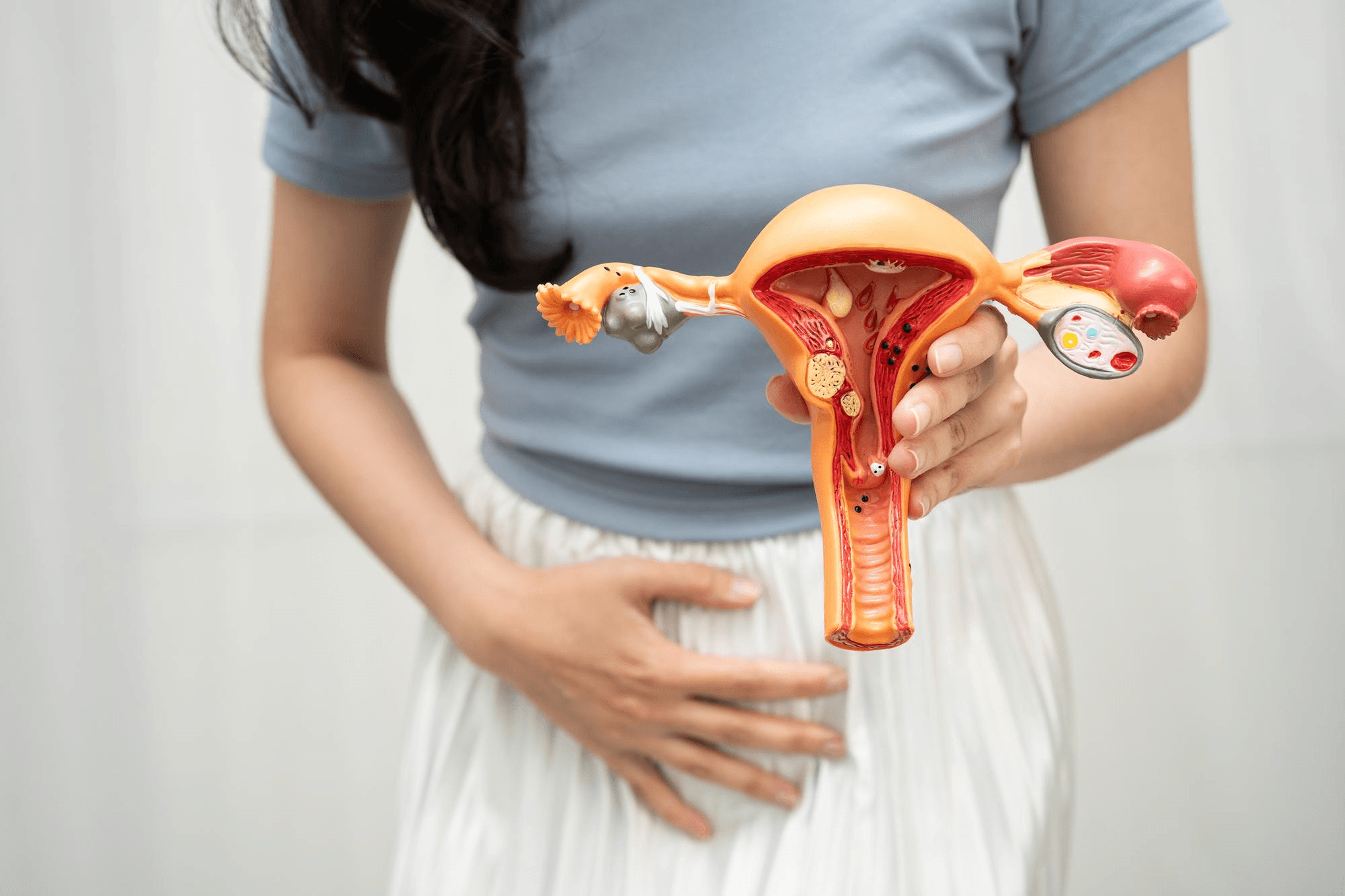
Medical illustration of the uterus showing multiple fibroids of different sizes, representing how untreated uterine fibroids can grow and affect reproductive health.
Heavy periods and a bulky, painful uterus are classic symptoms of both uterine fibroids and adenomyosis. The two conditions frequently coexist — and are sometimes confused with each other — but they are fundamentally different in what they are, how they are diagnosed, and what treatment works.
Dr. Shachi Singh, consultant gynaecologist and laparoscopic surgeon at Prakash Hospital, Sector 33, Noida, explains.
What Are Fibroids?
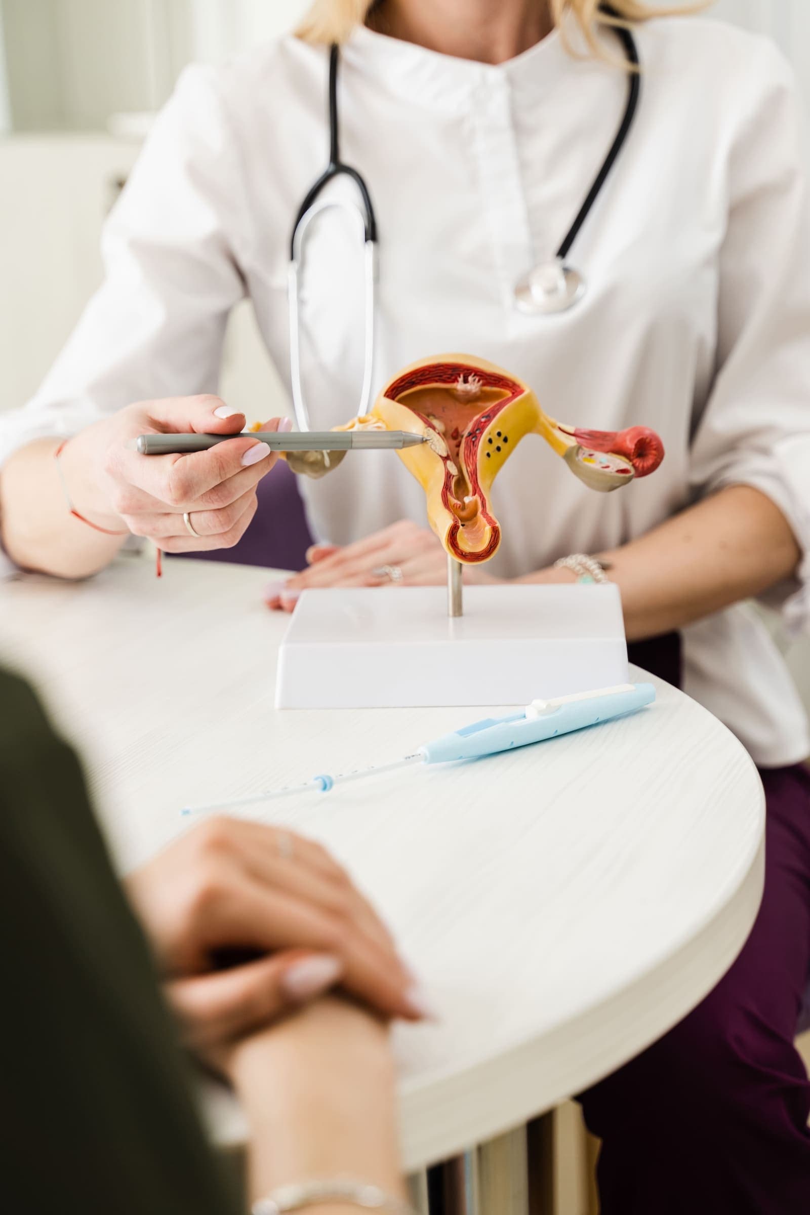
Fibroids (uterine leiomyomas) are benign tumours of the uterine smooth muscle. They are discrete, well-defined growths within or on the uterine wall — each fibroid is a separate entity with its own blood supply. They can be:
- Intramural: Within the uterine muscle wall (most common)
- Submucosal: Projecting into the uterine cavity — most symptomatic for bleeding and fertility
- Subserosal: Projecting from the outer uterine surface
- Pedunculated: Hanging on a stalk, either into the cavity or outside the uterus
Fibroids are oestrogen-dependent — they grow under oestrogen stimulation and typically stop growing (and often shrink) after menopause.
What Is Adenomyosis?
Adenomyosis is a completely different condition. In adenomyosis, endometrial glands and stroma (the tissue that normally lines the uterine cavity) grow into the uterine muscle wall itself. There are no discrete tumours — instead, endometrial tissue is scattered diffusely (or focally) throughout the myometrium.
This produces a uterus that is enlarged, soft, and globally thickened — described as "boggy" on examination. The ectopic endometrial tissue bleeds each month (responding to the same hormonal cycle as the normal endometrium), causing blood and inflammatory products to accumulate within the muscle, progressive uterine enlargement, and intense cramping.
Adenomyosis is also oestrogen-dependent. It is most common in women in their 30s and 40s, particularly those who have had pregnancies (the theory being that delivery disrupts the endometrial-myometrial junction). It improves after menopause.
How They Differ: Side by Side
| Feature | Fibroids | Adenomyosis |
|---|---|---|
| Nature | Discrete muscle tumours | Diffuse endometrial infiltration of muscle |
| Uterus feel | Irregular, firm, enlarged | Smooth, soft, uniformly enlarged |
| Main symptoms | Heavy periods, pressure, bulk symptoms | Heavy, painful periods, deep pelvic ache |
| Fertility impact | Submucosal fibroids most impactful | Can impair implantation and miscarriage risk |
| Diagnosis | Ultrasound (clear discrete lesions) | Ultrasound + MRI (more subtle features) |
| Response to menopause | Shrink | Resolves |
| Coexistence | Very common — up to 80% of adenomyosis patients also have fibroids |
Symptoms: Where They Overlap and Differ
Both cause heavy periods and an enlarged uterus. The distinctions:
Fibroids tend to cause:
- Heavy bleeding — particularly submucosal fibroids, which disturb the cavity and prevent normal uterine contraction to control bleeding
- Bulk symptoms from large fibroids: urinary frequency (pressure on the bladder), constipation, a visible abdominal mass
- Relatively less painful periods than adenomyosis
- Infertility if submucosal or large intramural
Adenomyosis tends to cause:
- Severe dysmenorrhoea — cramping pain that begins before the period and lasts throughout, often worse than fibroid-related pain
- Chronic deep pelvic aching between periods
- Painful intercourse (deep dyspareunia)
- Progressively worsening symptoms with each cycle
- Infertility — through impaired implantation and possibly increased miscarriage risk
Diagnosis
Ultrasound: Clear for fibroids — discrete, well-defined, hypoechoic lesions are easy to characterise by size, number, and location. Adenomyosis is subtler — features include globular uterine enlargement, asymmetrical myometrial thickening, heterogeneous (non-uniform) myometrial texture, myometrial cysts, and poorly defined lesions. An experienced sonographer can diagnose adenomyosis on ultrasound with reasonable accuracy.
MRI: The most accurate investigation for adenomyosis — shows the junctional zone (the inner myometrium) clearly. MRI also better characterises fibroids for surgical planning. Recommended when ultrasound findings are equivocal or when adenomyosis extent needs defining before treatment decisions.
Treatment
Fibroids
Medical management:
- Hormonal treatment (GnRH analogues, combined OCP, progestins, Mirena IUD) — reduces bleeding and fibroid size but effects are not permanent; fibroids return after treatment stops
- Tranexamic acid, NSAIDs — symptom management
Surgical management:
- Hysteroscopic myomectomy: for submucosal fibroids — removed via hysteroscope, no incisions
- Laparoscopic myomectomy: for intramural and subserosal fibroids — removes fibroids while preserving the uterus. Appropriate for women who want to keep fertility.
- Total laparoscopic hysterectomy (TLH): definitive — for women who have completed their family and want permanent resolution. No recurrence after hysterectomy.
- Uterine artery embolisation (UAE): radiological procedure occluding blood supply to fibroids — non-surgical option for selected women
Adenomyosis
Medical management:
- Mirena (levonorgestrel IUD): most effective non-surgical treatment for adenomyosis — reduces bleeding significantly and reduces adenomyosis-related pain in many women. First choice for women who want to preserve the uterus or fertility.
- Combined OCP (continuous use), progestin tablets, GnRH analogues: reduce oestrogenic stimulation and symptom severity. GnRH analogues are highly effective but produce temporary surgical menopause — not suitable long-term.
- Dienogest (a progestin): specific evidence for adenomyosis pain management
Surgical management:
- Hysterectomy: the only definitive cure for adenomyosis. Because adenomyosis is diffusely infiltrated throughout the muscle wall, it cannot be "removed" conservatively the way a fibroid can — the entire uterus must go. TLH (total laparoscopic hysterectomy) is the approach of choice for most women at Prakash Hospital. Complete and permanent resolution after hysterectomy.
- Adenomyomectomy: excision of focal adenomyosis — rarely performed, technically very challenging, high recurrence rate. Only appropriate in very selected cases when fertility preservation is paramount.
Gynaecological and Surgical Care in Noida and Greater Noida
Dr. Shachi Singh at Prakash Hospital, Sector 33, Noida, diagnoses and manages fibroids and adenomyosis — medically and surgically — for women across Noida and Greater Noida.
To book a consultation, call: +91 97023 46853
Clinic Hours: Monday to Saturday, 9 AM – 6 PM | Sunday, 10 AM – 2 PM
Address: D-12A, 12B, Sector-33, G.B. Nagar, Noida, UP 201301
This blog is for educational purposes only. Please consult Dr. Shachi Singh for assessment specific to your symptoms.