Breast Lump in Women: Causes, Fibroadenoma and When to Worry
Most breast lumps are benign, but every new lump should be examined promptly.
Finding a lump in your breast is frightening. Many women describe the same moment: a hand pauses in the shower or while dressing, there is something that was not there before, and the mind rushes straight to the worst possibility. Some women wait weeks before telling anyone, hoping it will disappear.
Please do not wait. Most breast lumps are not cancer. The majority turn out to be benign, such as fibroadenomas, cysts or hormonal changes. But the only way to know is to have the lump examined, because breast cancer is the most common cancer among women in India, and when it is found early, treatment is far more effective.
This guide explains the common causes of breast lumps, which features are reassuring and which are warning signs, how doctors evaluate a lump and what treatment involves. If you are looking for a gynaecologist in Noida to examine a lump or arrange further tests, details are at the end.
Why breasts feel lumpy
The breast is made up of glandular tissue (lobules that produce milk and ducts that carry it), fat and fibrous connective tissue. Under the influence of hormones through the menstrual cycle, the glands swell and soften, and many women notice their breasts becoming lumpy, heavy or tender before their periods and settling afterwards. This "cyclical lumpiness" is normal. Breast tissue is naturally nodular, and what feels like a lump may be the edge of a normal structure like the ribs or a ridge of tissue at the lower margin of the breast.
The key is to know your own breasts, so you can notice what is new, different or persistent.
Common causes of breast lumps
1. Fibroadenoma
The most common benign breast tumour in young women, typically between 15 and 35 years. A fibroadenoma is a firm, smooth, rubbery, round or oval lump that moves easily under the skin, which is why it is sometimes called a "breast mouse." It is usually painless, grows slowly and does not change with the menstrual cycle. It is made up of glandular and connective tissue, and is not cancer. Some grow during pregnancy or breastfeeding and shrink after menopause. Several can occur in one or both breasts.
2. Breast cysts
Fluid-filled sacs, common in women aged 30 to 50, especially before menopause. They feel like a smooth, round or oval lump, may be tender and can change in size with the menstrual cycle. Ultrasound easily distinguishes a simple cyst from a solid lump. Cysts are benign, and large painful ones can be drained with a needle.
3. Fibrocystic changes
A common, benign condition in which breast tissue becomes lumpy, thickened or ropey, often with tenderness or pain that varies with the cycle. It is not a disease, and does not usually need treatment other than reassurance and supportive measures.
4. Infection and inflammation
- Mastitis: inflammation, commonly in breastfeeding women, with pain, redness, warmth, swelling and sometimes fever.
- Breast abscess: a collection of pus, causing a painful, hot, tender lump, which needs drainage and antibiotics.
5. Fat necrosis
A firm lump that follows an injury or surgery, caused by damaged fatty tissue. It may look worrying on imaging and sometimes needs a biopsy to confirm.
6. Lipoma
A soft, movable fatty lump, harmless.
7. Galactocele
A milk-filled cyst, often in or after breastfeeding.
8. Intraductal papilloma
A small wart-like growth inside a milk duct, which can cause a lump and bloody or clear nipple discharge. It is usually benign but needs evaluation.
9. Phyllodes tumour
An uncommon tumour that can grow quickly, usually benign, but can be borderline or malignant. It generally requires surgical removal.
10. Breast cancer
Cancer may present as a lump, though it can also present without one. Cancer is more common with age, but can occur in younger women too. Features that raise concern are described below.
Other causes
Lymph nodes in the armpit (axilla) swollen by infection or other conditions can be felt as lumps. Breast changes in men (gynaecomastia) also occur but are outside the scope of this article.
Features: reassuring vs concerning
Appearance and feel can't tell benign from malignant with certainty, so every new lump needs assessment. Still, certain features guide the level of concern.
More reassuring features:
- Smooth, round or oval
- Soft or rubbery
- Moves easily under the skin
- Tender and changing with the menstrual cycle
- Present in a younger woman
- Known to have been stable for a long time
Features that raise concern:
- A hard, irregular or fixed lump, which feels stuck to the skin or chest wall
- A lump that is growing or has persisted through a full menstrual cycle
- Skin changes: dimpling or puckering, an "orange peel" texture, redness, thickening, or an ulcer
- Nipple changes: turning inward (new inversion), scaling, crusting or a rash that doesn't heal
- Nipple discharge that is bloody, or comes from one nipple spontaneously
- A lump in the armpit or swelling above the collarbone
- Change in the size or shape of the breast
- A lump after menopause
- A family history of breast or ovarian cancer
- Persistent pain in one spot (less common as a presenting sign, but should be evaluated)
Painless lumps are not necessarily safe. Many cancers are painless, so absence of pain isn't reassuring.
When to see a doctor
See a doctor promptly, ideally within a week or two, if you notice:
- Any new lump or thickening in the breast or armpit
- A lump that persists after your next period
- Any of the concerning features above
- Nipple discharge that is bloody or occurs without squeezing
- Skin or nipple changes
- A breast lump during pregnancy or breastfeeding that doesn't settle in a week or two with treatment
Do not wait for a "convenient" time. Early evaluation brings peace of mind in most cases, and early diagnosis in the few that need it.
How is a breast lump evaluated?
Doctors use a standard approach, often called triple assessment, combining three elements to reach a reliable diagnosis.
1. Clinical examination and history
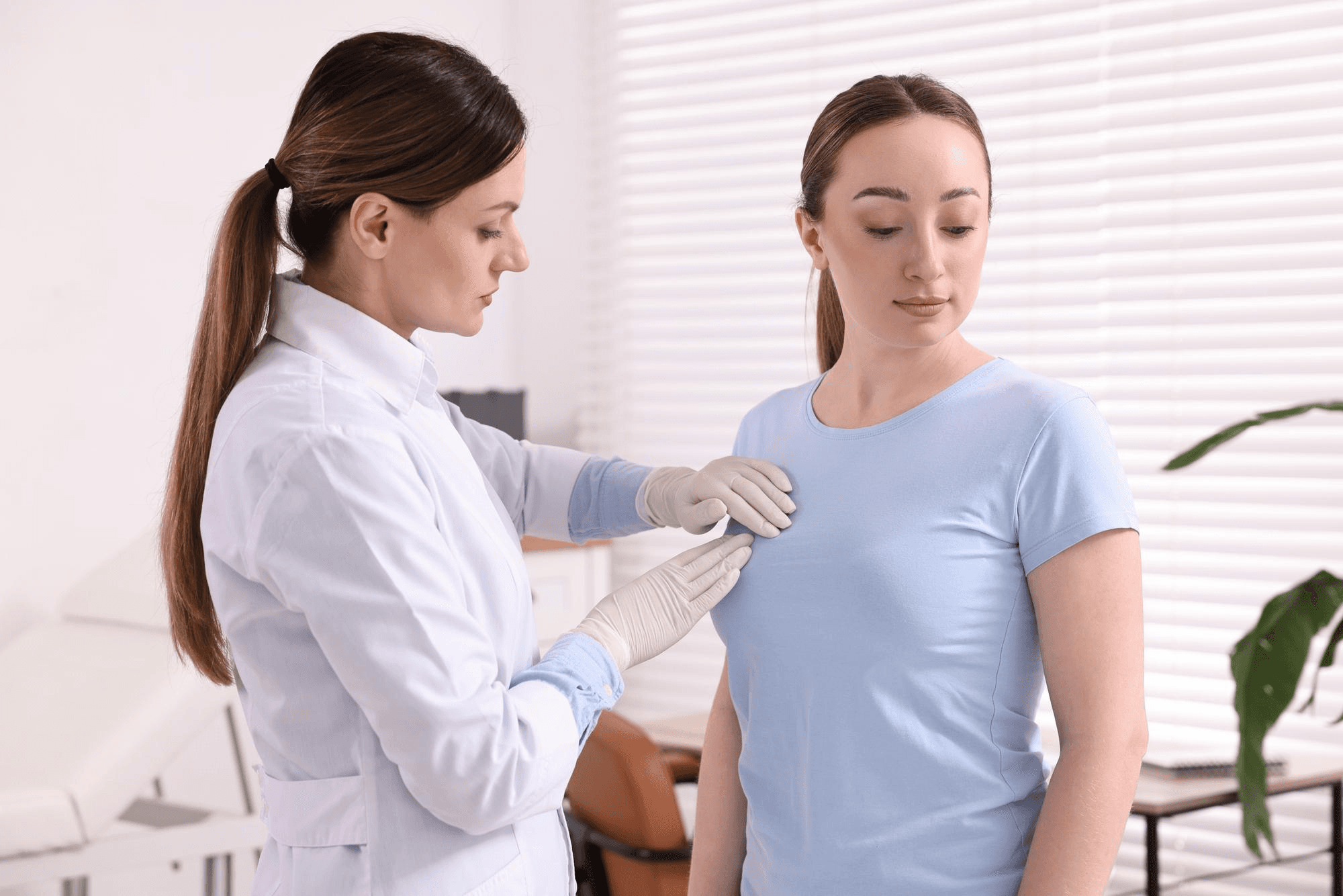
I ask about when you noticed the lump, any change over time, pain, discharge, your periods, pregnancies, breastfeeding, hormone use, previous breast problems and family history of breast, ovarian or other cancers. I examine both breasts and the armpits, with you sitting and lying down, in a private setting. A chaperone can be present if you wish.
2. Imaging
- Ultrasound is the first test for women under 30 to 40, because young breasts are dense and mammography is less useful. It can tell a cyst from a solid lump, and characterise a solid lump.
- Mammography (X-ray of the breast) is used for women over 40, and sometimes younger when indicated. It detects small cancers and microcalcifications. Mammography and ultrasound are often used together.
- MRI in selected cases: high-risk women, unclear findings or extent of disease.
3. Tissue sampling
If a lump looks suspicious or can't be clearly classified, a sample is taken:
- Fine-needle aspiration cytology (FNAC): a thin needle removes cells for examination. It's quick and useful for cysts and some lumps.
- Core needle biopsy: a slightly larger needle takes a small cylinder of tissue, providing a more definite diagnosis and tumour characteristics. It's performed under local anaesthesia, usually with ultrasound guidance.
- Excision biopsy: surgical removal of the lump for examination when needed.
Together these give an accurate diagnosis in the great majority of women.
Treatment
Treatment depends entirely on the diagnosis.
Simple cyst: reassurance; aspiration if large, painful or if the diagnosis is uncertain. Recurrence is possible.
Fibroadenoma: often just monitored with ultrasound, especially in young women when it is small and confirmed by biopsy. Surgical removal may be advised if it is large (typically over 3 to 5 cm), growing, causing discomfort or the diagnosis is uncertain, or if you prefer removal. Newer techniques such as vacuum-assisted excision can remove some fibroadenomas through a tiny cut.
Fibrocystic changes: reassurance, a supportive well-fitting bra, reducing caffeine if it helps (evidence is limited), pain relief when needed and sometimes medicines in severe cases.
Infection/abscess: antibiotics, drainage when needed, and continuing breastfeeding if possible.
Fat necrosis, lipoma, papilloma: often observed or removed if symptomatic or uncertain.
Phyllodes tumour: surgical removal with adequate margins.
Breast cancer: treatment is individualised and may involve surgery (breast-conserving surgery or mastectomy), radiotherapy, chemotherapy, hormone therapy or targeted therapy, delivered by a multidisciplinary team. Early-stage breast cancer has an excellent outlook, which is why early detection matters so much.
If a concern for cancer arises, I would arrange prompt referral to a breast surgeon or oncology team for definitive treatment, and support you throughout.
Breast awareness and screening
Regular attention to your breasts can help find problems early.
- Be breast aware. Know how your breasts normally look and feel at different times in your cycle. Look and feel for changes. A formal monthly "self-examination" has not been proven to reduce deaths, but awareness and reporting changes promptly is strongly encouraged.
- Clinical breast examination by a doctor, as part of regular check-ups.
- Mammography is generally recommended from about 40 years (some guidelines suggest 45 or 50), at intervals your doctor will advise, and earlier for women with a strong family history.
- Genetic counselling if you have a family history of breast or ovarian cancer, young-onset cancer or a known BRCA mutation.
- Healthy lifestyle: maintain a healthy weight, be physically active, limit alcohol, avoid smoking, and breastfeed if you can, all of which are linked to lower risk.
Breast lumps in pregnancy and breastfeeding
Breasts change dramatically in pregnancy and lactation, and benign lumps such as galactoceles and fibroadenomas are common. Still, any new lump in pregnancy or breastfeeding should be examined. Ultrasound is safe, and biopsy can be done. Pregnancy-associated breast cancer is rare but real, and delay in diagnosis is common because lumps are assumed to be harmless.
What you can do at home while waiting for your appointment
- Don't squeeze or massage the lump.
- Note its size, position, whether it changes with your period and any other changes.
- Avoid unproven creams or herbal applications.
- Try not to catastrophise. Most lumps are benign, and a calm approach helps.
- Bring a family member or friend for support, if you like.
Common questions
Does a painful lump mean it's not cancer? Pain makes cancer less likely but doesn't rule it out.
Can a fibroadenoma turn into cancer? The risk is very low, though some complex fibroadenomas are associated with a slightly increased risk over time.
Do I need a mammogram at 25? Not usually. Ultrasound is the preferred first test in young women.
Can a bra cause breast cancer? No.
Does a breast lump mean I can't breastfeed? Most benign lumps do not affect breastfeeding.
Can stress cause breast lumps? Stress itself doesn't cause lumps, though it can make you more aware of breast discomfort.
Should I get any new lump checked even if I had a normal mammogram? Yes. A normal mammogram does not exclude all cancers, and a new lump needs clinical and imaging assessment.
Get any new lump examined
A prompt check is the safest path, and in most cases ends in reassurance. I see women from Noida, Greater Noida, Ghaziabad and Delhi NCR at Prakash Hospital, Sector 33, and will arrange imaging and biopsy or referral if required. To book a consultation, call +91 97023 46853.
Related reading
- Breast self-examination: how to do it
- BRCA mutations and breast cancer risk
- Women's health after 40
- Mastitis while breastfeeding
- Pap smear test: when and why
This article is for general education. Any new breast lump should be examined by a doctor.