Molar Pregnancy: Symptoms, Diagnosis, Treatment and Follow-Up

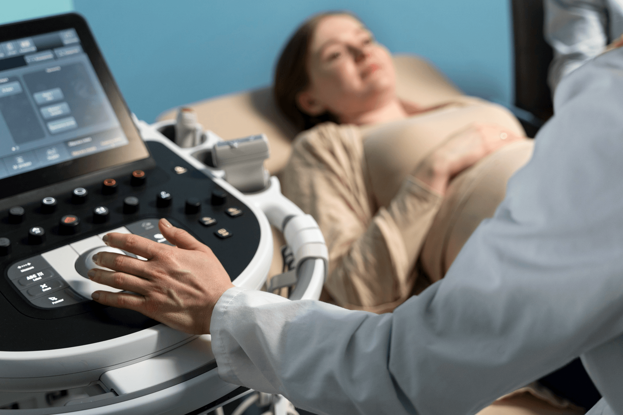
Ultrasound is the key test; a complete mole often shows a snowstorm pattern and no fetus.
A molar pregnancy is one of the most distressing surprises that can happen in early pregnancy. A woman sees a positive test, perhaps feels very nauseous, and then an ultrasound shows "something unusual." The word "mole" or "molar" is mentioned, and suddenly the happiness of a new pregnancy is replaced by confusion, fear and grief.
I want to begin with some reassurance. A molar pregnancy is not caused by anything you did or did not do. It is a rare abnormality of fertilisation, and in the great majority of women it is treated successfully and does not affect their ability to have healthy babies in the future. It does need careful follow-up, which is the main part of the management.
This guide explains what a molar pregnancy is, its types and causes, the symptoms, how it's diagnosed and treated, why follow-up with hCG tests matters and what to expect afterwards. If you want to discuss your own situation with a gynaecologist in Noida, details are at the end.
What is a molar pregnancy?
A molar pregnancy, also called a hydatidiform mole, is a type of gestational trophoblastic disease (GTD). It happens when a pregnancy does not develop normally right from fertilisation. Instead of forming a healthy placenta and a baby, the cells that should become the placenta (trophoblast cells) grow abnormally and rapidly, creating a mass of small fluid-filled sacs that look like a bunch of grapes on ultrasound or under the microscope.
A molar pregnancy cannot result in a living baby. The pregnancy cannot continue, and it must be removed. It is not a cancer, but in some cases the abnormal tissue can persist or invade the uterus after treatment, so follow-up is vital.
Types of molar pregnancy
Complete mole
An egg with no genetic material from the mother is fertilised by one sperm (which duplicates its DNA) or two sperm. The result is a set of chromosomes entirely from the father. There is no embryo or fetal tissue, only abnormal placental tissue that fills the uterus. Complete moles tend to grow quickly, produce very high levels of hCG and carry a higher risk of persistent disease (about 15 to 20 percent).
Partial mole
A normal egg is fertilised by two sperm, or one sperm that duplicates, giving a cell with three sets of chromosomes (triploidy). There may be some embryonic or fetal tissue, but it is abnormal and cannot survive. The placental changes are patchy. Partial moles usually produce lower hCG and have a lower risk of persistent disease (about 1 to 5 percent).
How common is it?
Molar pregnancy is uncommon. In Western countries, it occurs in about 1 in 1,000 pregnancies. It is more common in parts of Asia, including India, with some studies reporting higher rates, though estimates vary. Risk factors include:
- Age: very young (under 16 to 20) or older (over 35 to 40), with the highest risk in women over 45
- A previous molar pregnancy: the risk of another is higher (about 1 to 2 percent)
- Previous miscarriages
- Possibly dietary factors and nutritional deficiencies (under study)
- Family history in rare genetic forms
Most women who have a molar pregnancy have none of these risk factors.
Symptoms
A molar pregnancy can look much like an ordinary early pregnancy at first, with a missed period and a positive test. The signs that suggest something is different include:
- Vaginal bleeding in the first trimester (the most common symptom), often dark brown or bright red, and sometimes with watery discharge. Occasionally, small grape-like vesicles are passed.
- Severe nausea and vomiting (hyperemesis), often more than expected, due to very high hCG
- A uterus larger than expected for the dates
- Pelvic pain or pressure
- Very high hCG levels on blood tests
- Signs of an overactive thyroid (rapid pulse, sweating, tremor), because hCG can stimulate the thyroid
- High blood pressure or preeclampsia-like signs before 20 weeks, which is unusual and a red flag
- Anaemia from ongoing bleeding
- Ovarian cysts (theca lutein cysts) due to high hCG
- Rarely, breathlessness if the tissue spreads
Some women have no symptoms, and the diagnosis comes from a routine early scan.
How is it diagnosed?
Ultrasound
The key investigation. A complete mole classically appears as a "snowstorm" or "cluster of grapes" pattern, with a uterus filled with mixed echoes and small cystic spaces, and no fetus. A partial mole may show a thickened, cystic placenta with an abnormal fetus or an empty-looking sac. Early in pregnancy, the picture can be subtle, and a molar pregnancy may be mistaken for a missed or incomplete miscarriage.
Blood tests
- Serum beta-hCG: usually much higher than expected for the gestational age, especially in complete moles
- Complete blood count, blood group and Rh factor
- Thyroid function tests, liver and kidney function
- Chest X-ray in selected cases, if there are symptoms or high hCG, to look for spread
Pathology
The tissue removed at evacuation should always be sent for histopathology (examination under the microscope). This confirms the diagnosis and distinguishes complete from partial mole. Special tests may be needed in uncertain cases.
This is why every pregnancy tissue removed after a miscarriage, particularly missed miscarriage, should be sent for examination.
Treatment
Evacuation of the uterus
The standard treatment is suction evacuation (suction curettage), done under anaesthesia in a hospital. A thin tube attached to a gentle suction device removes the abnormal tissue from the uterus. It is preferred to medical methods or sharp curettage, as it lowers the risk of bleeding, perforation and incomplete removal. Oxytocin may be given during or after the procedure to help the uterus contract. If you are Rh negative, you will usually receive anti-D immunoglobulin.
Medicines to induce evacuation (like misoprostol) are generally avoided, because they can increase bleeding and the risk of persistent disease.
Hysterectomy
Rarely, in women who have completed their family and are older, hysterectomy may be considered. This removes the uterus but does not eliminate the need for hCG follow-up.
Treating complications
High blood pressure, thyroid overactivity, anaemia and severe vomiting are treated as required.
Follow-up: the most important part
After evacuation, the aim is to make sure that no abnormal tissue remains or regrows. This is done by monitoring hCG levels in the blood (and sometimes urine).
- Frequency: hCG is measured weekly until it falls to normal (undetectable) levels, usually within 8 to 12 weeks. After that, monitoring continues with monthly tests.
- Duration: after a complete mole, follow-up typically continues for about 6 months after hCG normalises (in many protocols); for a partial mole, shorter follow-up, often a few more tests after the first normal result, in line with current guidelines. Your doctor or specialist centre will advise a schedule.
- What is expected: a steady fall in hCG. If hCG plateaus (stays the same), rises or doesn't fall as expected, it suggests persistent trophoblastic disease.
In some centres, follow-up is coordinated by a specialist trophoblastic disease unit. Ask your doctor where your hCG results are tracked and who will review them.
Contraception during follow-up
You must avoid pregnancy during follow-up, because a rise in hCG from a new pregnancy cannot be distinguished from regrowth of the mole. Usually, advice is to use reliable contraception for the entire follow-up period (for example about 6 to 12 months for complete moles), as directed. The combined oral contraceptive pill is commonly used and is safe once hCG is normal, and other methods such as condoms, hormonal implants and injections can be used. Your doctor will advise on the best choice. Intrauterine devices are usually avoided until hCG is normal because of the risk of perforation if the tissue persists.
Persistent trophoblastic disease (GTN)
In a minority of women, abnormal tissue persists or grows after evacuation. This is called gestational trophoblastic neoplasia (GTN). It is diagnosed when hCG levels plateau, rise or stay elevated after evacuation. Tests (scans and sometimes imaging of the chest or brain) check for spread.
The good news is that GTN is among the most curable of cancers. Most women are treated with chemotherapy, typically methotrexate or actinomycin D as single agents for low-risk disease, and cure rates exceed 95 to 98 percent. Higher-risk disease requires combination chemotherapy at a specialist centre and still has very high cure rates. Treatment preserves fertility in most cases. Hysterectomy is rarely required.
Emotional impact
A molar pregnancy is a loss. You may grieve a pregnancy that felt real, while also dealing with the medical terminology, the need for tests and the delay before you can try again. Feelings of guilt, anger or sadness are common. You are not to blame.
- Talk to your partner, a close friend or a counsellor
- Ask your doctor any questions, however small
- Remember that follow-up is a precaution, and the outlook is generally excellent
- Give yourself time before the next pregnancy
Future pregnancies
Most women who have had a molar pregnancy go on to have healthy, normal pregnancies. The chance of another molar pregnancy is small, about 1 to 2 percent, but higher than for women who haven't had one. In a future pregnancy, you should:
- Have an early ultrasound (first trimester) to confirm normal development
- Have an hCG level checked about 6 to 8 weeks after the end of the next pregnancy (whether it ends in birth, miscarriage or termination) to make sure no tissue has recurred
- Send any miscarriage tissue for pathology
Wait until your follow-up is complete and your doctor gives the go-ahead before trying again.
When to see a doctor urgently
- Heavy vaginal bleeding after evacuation
- Persistent or increasing pelvic pain
- Fever
- Breathlessness, cough or chest pain
- Severe headache, visual disturbances or seizures
- A positive pregnancy test when you should be using contraception during follow-up
Common questions
Is a molar pregnancy cancer? No, but a small proportion can turn into a persistent tumour needing treatment, which is why follow-up is essential.
Can I have a normal pregnancy afterwards? Yes, in the vast majority of cases.
Could I have prevented it? No.
Will it affect my fertility? Not generally.
Is chemotherapy always needed? No. Only a minority of women need it.
How long do I need to wait before trying again? Usually until follow-up is complete, as advised by your doctor, commonly six months to a year depending on the type and hCG course.
Can I still breastfeed or have symptoms like a normal pregnancy? In molar pregnancy, no baby is born, so there's no breastfeeding. Symptoms settle after evacuation.
Support when you need it
A molar pregnancy is rare, and good follow-up makes all the difference. I look after women from Noida, Greater Noida, Ghaziabad and Delhi NCR at Prakash Hospital, Sector 33, and I make sure every patient understands the follow-up plan. To book a consultation, call +91 97023 46853.
Related reading
- Ectopic pregnancy warning signs
- Recurrent miscarriage: causes and tests
- Hyperemesis gravidarum
- Missed miscarriage: signs and management
- Early pregnancy symptoms
This article is for general education. Follow-up of a molar pregnancy must be guided by your own doctor.