Endometrial Hyperplasia: Understanding the Diagnosis and What Comes Next
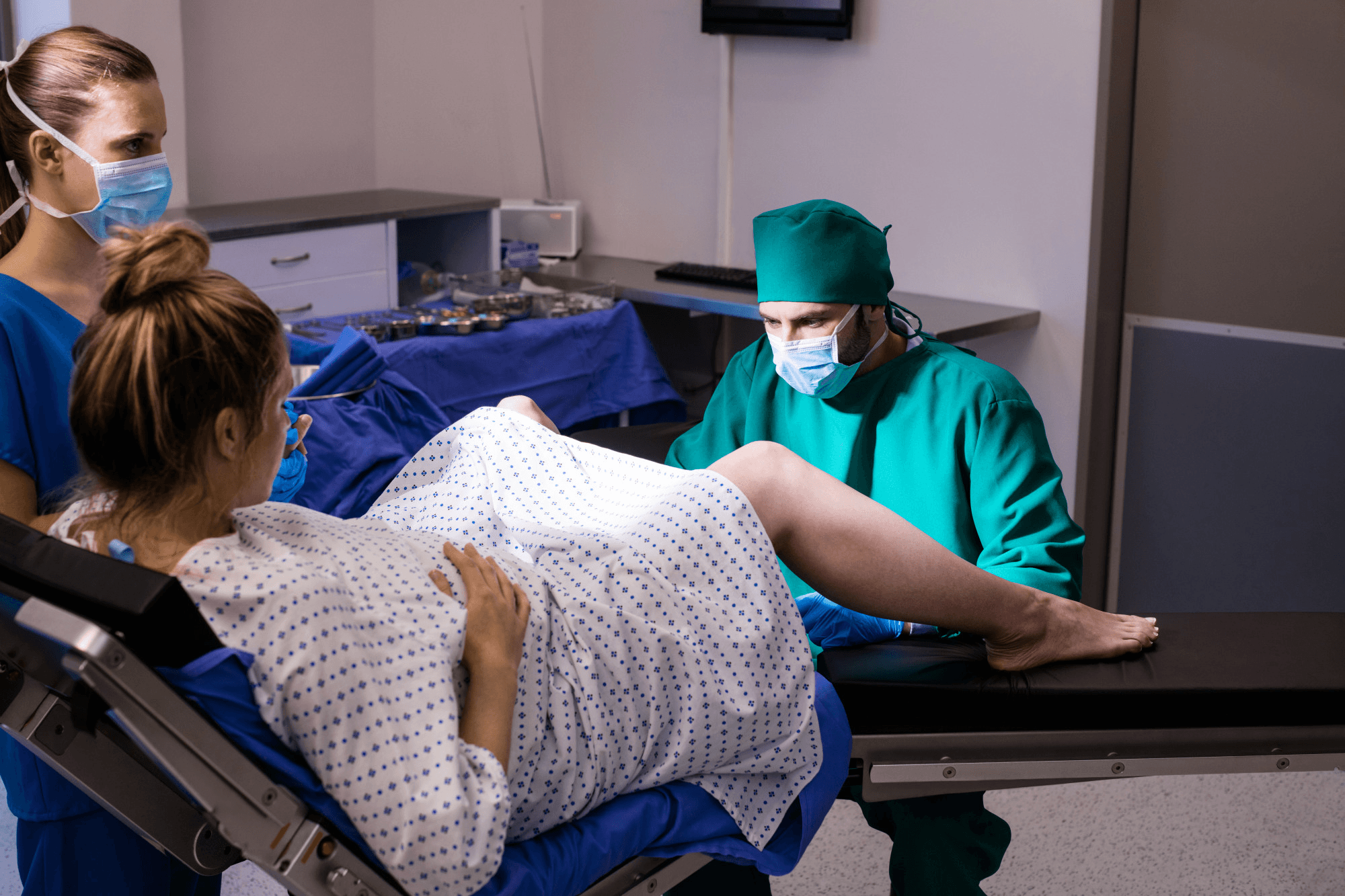
A medical team of doctors conducts laparoscopic surgery in an operating room where they use specialised equipment to remove lesions. And make a woman fertile by treating her endometriosis.
Endometrial hyperplasia — an abnormal thickening of the uterine lining — is a diagnosis that ranges from a benign, easily managed condition to a significant precursor to endometrial cancer, depending on its subtype. Understanding which type a woman has is the essential first step, because it completely determines the management pathway.
Dr. Shachi Singh, consultant gynaecologist at Prakash Hospital, Sector 33, Noida, explains.
What Endometrial Hyperplasia Is
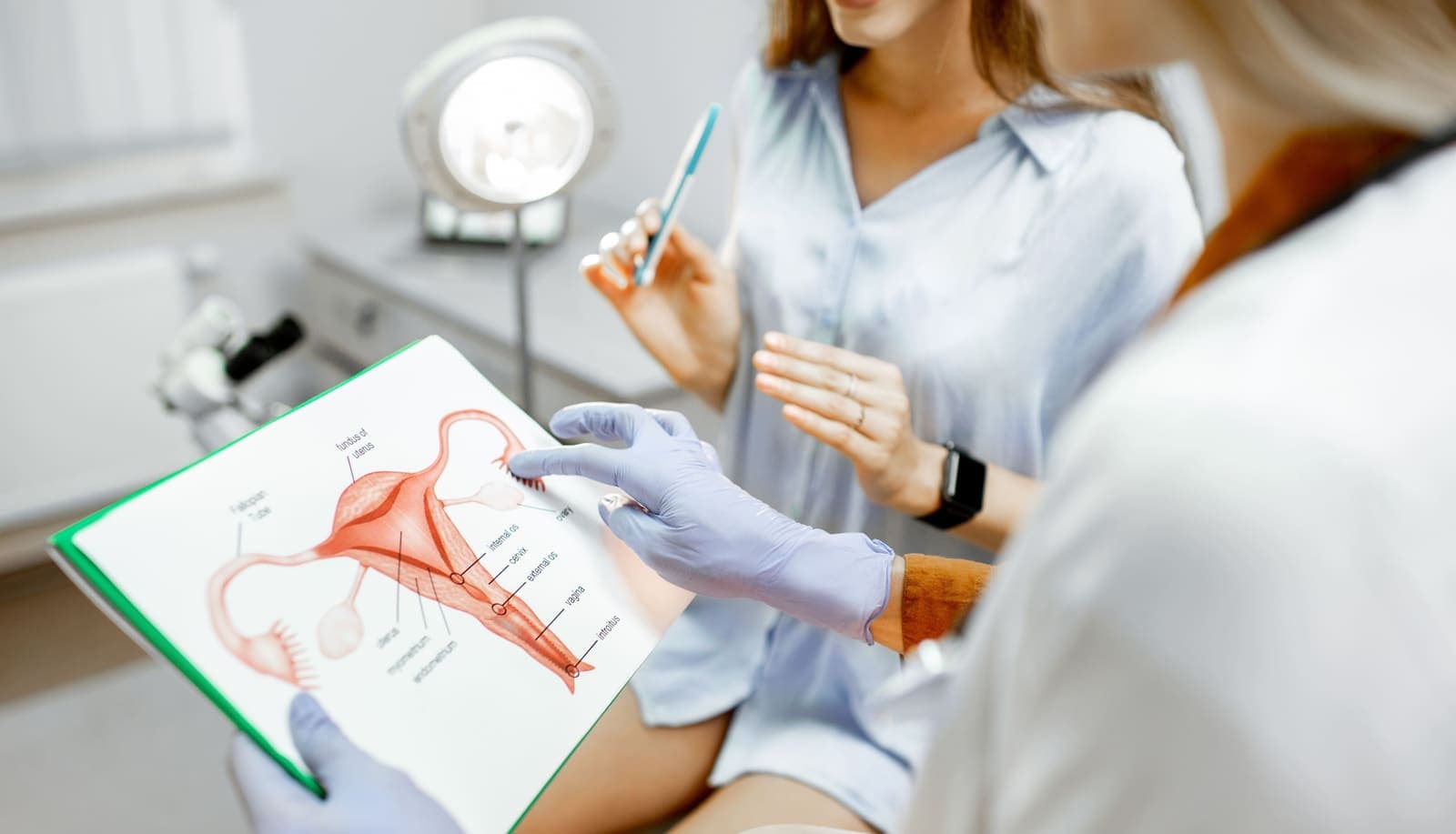
The endometrium (uterine lining) normally thickens in the first half of the menstrual cycle under oestrogen stimulation, then sheds at menstruation. When progesterone is absent or insufficient — or when oestrogen stimulation is prolonged or excessive — the endometrium does not shed normally and instead undergoes abnormal proliferation (thickening).
This abnormal proliferation is endometrial hyperplasia. The endometrial glands become crowded, distorted, and increased in number. Depending on whether the individual cells show normal or abnormal appearance (atypia), the condition has very different implications.
Types: With and Without Atypia
Endometrial hyperplasia without atypia (simple or complex):
The glands are crowded and increased in number, but the individual cells appear normal microscopically. The risk of progression to endometrial cancer is low — approximately 1 to 3% over 10 to 20 years. This type responds well to progestin treatment and in many cases regresses spontaneously.
Endometrial hyperplasia with atypia (atypical hyperplasia):
The glands are crowded and the individual cells show abnormal features (nuclear enlargement, irregular chromatin, prominent nucleoli). This is a significant premalignant lesion. The risk of concurrent endometrial cancer found at hysterectomy in women diagnosed with atypical hyperplasia on biopsy is approximately 25 to 40% — meaning the biopsy may have missed an area of cancer. The risk of progression to cancer if untreated is approximately 25 to 30% over years. This type requires more aggressive management.
Who Is at Risk
Risk is related to prolonged or excessive oestrogen exposure relative to progesterone:
- Obesity: Adipose tissue produces oestrogen independently of the ovaries. Obese women have chronically elevated oestrogen without corresponding progesterone from ovulation.
- PCOS: Chronic anovulation (failure to ovulate) means prolonged oestrogen exposure without cyclical progesterone from the corpus luteum.
- Late menopause (after 55): Prolonged reproductive years of oestrogen exposure.
- Nulliparity (never having been pregnant): Pregnancy provides prolonged progesterone exposure. Women who have never been pregnant lack this protective effect.
- Tamoxifen: Used for breast cancer treatment, tamoxifen has oestrogen-like effects on the uterine lining.
- Oestrogen-only HRT: Post-menopausal oestrogen therapy without progestogen causes endometrial stimulation. This is why combined HRT (oestrogen + progestogen) is used in women with an intact uterus.
- Diabetes and insulin resistance: Increases oestrogen production through multiple mechanisms.
- Lynch syndrome: An inherited condition affecting mismatch repair genes — dramatically increases lifetime risk of endometrial (and colorectal) cancer.
Symptoms
Irregular or heavy vaginal bleeding — the most common presentation. In premenopausal women: prolonged or heavy periods, irregular periods, bleeding between periods. In postmenopausal women: any vaginal bleeding (which should always be investigated).
No symptoms — endometrial hyperplasia is occasionally found incidentally on ultrasound (thickened endometrium) or during investigation of other gynaecological conditions.
Diagnosis
Transvaginal ultrasound: Shows a thickened endometrium — in postmenopausal women, the endometrial thickness threshold prompting biopsy is typically 4 to 5 mm. In premenopausal women, the normal thickness varies across the cycle.
Endometrial biopsy: The definitive diagnostic test. A thin tube is passed through the cervix to obtain a small sample of endometrial tissue. Usually done in the clinic under local anaesthesia. Provides tissue for histological examination — identifying whether hyperplasia is present and whether atypia is present.
Hysteroscopy with directed biopsy: More complete sampling of the uterine cavity. Preferred when the biopsy sample is insufficient or when a focal lesion is seen on ultrasound.
Treatment
Hyperplasia without atypia:
Progestin treatment: Progesterone suppresses endometrial growth. Options include:
- Levonorgestrel-releasing IUS (Mirena) — the most effective progestin delivery system for endometrial hyperplasia. Delivers progestin directly into the uterine cavity at consistently high local concentrations. Complete regression rates of 90 to 96% at 12 months.
- Oral progestins: Medroxyprogesterone acetate, norethisterone, or micronised progesterone. Effective but require consistent daily compliance.
Follow-up endometrial biopsy at 6 to 12 months confirms regression. If regression is confirmed, ongoing surveillance is maintained.
Addressing modifiable risk factors: Weight loss in obese women significantly reduces oestrogen levels and improves response to progestin treatment. Treating PCOS, managing diabetes.
Hyperplasia with atypia:
For women who have completed their family: hysterectomy is the recommended treatment. The significant concurrent cancer risk (25 to 40%) and the high progression risk make uterine conservation risky. Total laparoscopic hysterectomy is the preferred approach where feasible.
For women who wish to preserve fertility: High-dose progestin treatment (Mirena plus oral progestin) under close surveillance, with biopsy every 3 to 6 months. This is a non-standard approach requiring careful individualised discussion, specialist input, and the understanding that hysterectomy remains the definitive treatment. Any atypical hyperplasia that does not regress on progestin treatment requires hysterectomy.
Gynaecological Care in Noida and Greater Noida
Dr. Shachi Singh at Prakash Hospital, Sector 33, Noida, diagnoses and manages endometrial hyperplasia — including endometrial biopsy, Mirena insertion, and laparoscopic hysterectomy — for women across Noida and Greater Noida.
To book a consultation with Dr. Shachi Singh, call: +91 97023 46853
Clinic Hours: Monday to Saturday, 9 AM – 6 PM | Sunday, 10 AM – 2 PM
This blog is for informational purposes only. Please consult Dr. Shachi Singh or a qualified gynaecologist for assessment specific to your situation.